Hidden 3D Genome Loops That Survive Cell Division

Gentaur
29 Oct 2025
Recent work from Massachusetts Institute of Technology (MIT) challenges long-standing views of how the genome is organized during cell division. For decades, the field assumed that most three-dimensional (3D) genome structure collapses when a cell enters mitosis and then rebuilds after division. However, MIT researchers show that small, highly specific loops of DNA, connecting regulatory elements like enhancers to genes persist during mitosis and may even strengthen.
What we thought we knew ?
Genome architecture in interphase
In a non-dividing (interphase) cell, the genome folds into a complex 3D structure. DNA is packaged in the nucleus, and within that packaging you find loops, compartments, and domains. These enable genes to interact with regulatory regions (enhancers, promoters) that may be far away in linear sequence.
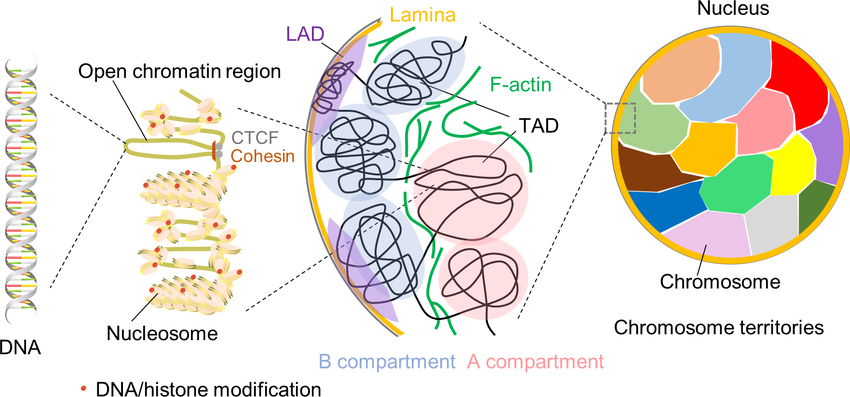
For example, the method Hi‑C (Wikipedia) enabled mapping of DNA contacts across the genome. Loops and domains (such as topologically associating domains, or TADs) are believed to support regulatory programmes that define cell identity, gene expression patterns, and so on.
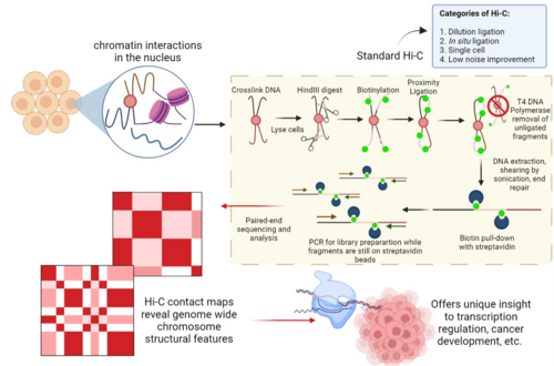
What we believed happens during mitosis ?
When a cell enters mitosis, chromosomes become highly compacted. The classical view was: large-scale genome architecture (loops, TADs, A/B compartments) falls apart. Transcription largely shuts down. After division, re-establishment of the 3D structure allows the gene regulatory network to restart.
Thus, mitosis was thought to be a “reset” of genome architecture a blank slate, so to speak.
The New Study: What MIT Found ?
A high-resolution technique
The MIT team used a method called Region‑Capture Micro‑C (RC‑MC) (NCBI). RC-MC is a refinement over Hi-C: it employs a different enzyme that cuts the genome into smaller, more uniform fragments; it uses capture of selected genome regions; and it attains 100 to 1,000× higher depth in target regions compared to classic Hi-C in some settings.
This enhanced resolution enabled the team to detect finer structures that earlier methods missed.
Discovery of “microcompartments”
By applying RC-MC, the researchers identified small structures they call microcompartments (Wikipedia) : dense loops connecting nearby enhancers and promoters (regulatory DNA) that form local clusters of interaction.
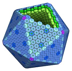
Interestingly, these microcompartments behave differently from the larger loops, TADs and compartments. The mechanism forming them appears distinct.
Persistence through mitosis
The key new finding is that these microcompartments persist during mitosis, when chromosomes are highly compacted. More surprisingly, they appear to strengthen during cell division (especially in anaphase/telophase) before weakening again in the G1 phase.
In contrast, the large-scale structures (A/B compartments, TADs) behave as expected: they disappear during mitosis.
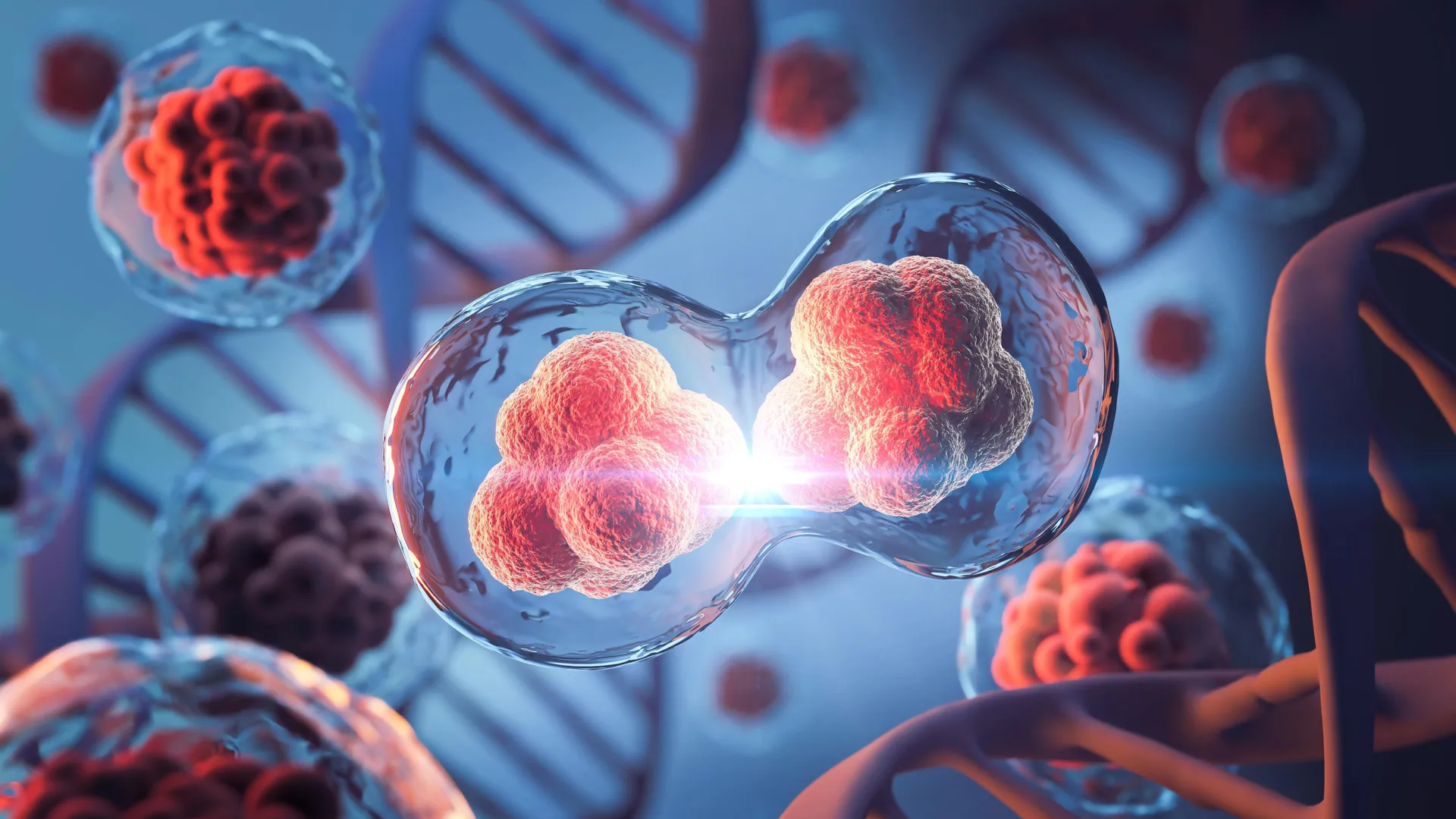
A new genome-mapping method revealed that small DNA loops persist and intensify during mitosis, challenging the belief that genome structure dissolves during division. These “microcompartments” may drive brief bursts of transcription and help cells preserve regulatory memory. Credit : Shutterstock
A potential functional link to transcription
The team also observed that these microcompartments are enriched near genes that show a brief spike of transcription during mitosis or at mitotic exit. That spike has puzzled researchers for decades. They propose that as chromosomes compact, enhancers and promoters are brought into closer proximity, which fosters loop formation and may trigger unintended transcription bursts. Then, once the cell enters G1, many loops are pruned.
Detailed Mechanistic Insights
Why do microcompartments survive and even strengthen?
Based on the study and commentary :
- During mitosis, chromatin compaction is very high. This reduces nuclear volume and pushes DNA regions closer together.
- Regulatory elements (enhancers/promoters) that might be far apart in linear distance come closer in 3D. This proximity promotes loop formation (or strengthens pre-existing loops).
- The “stickiness” or affinity between certain regulatory elements might favour formation of stable small loops under these compact conditions. The authors suggest compaction + homotypic affinity = microcompartment formation.
- After mitosis, the cell enters G1. At this point, the chromatin de-compacts, and the cell may actively prune many of these loops to restore the regulated architecture of interphase.
How this links structure to function ?
Because microcompartments connect regulatory DNA and genes, their survival through mitosis means that some memory of regulatory architecture is preserved across cell division. This may help daughter cells more quickly restore regulatory programmes. The usual view was that everything resets; this study suggests that at least some genome memory persists.
The observation of transcriptional spike: Genes close to microcompartments show elevated transcription at the end of mitosis. This could reflect accidental activation due to physical proximity of regulatory elements caused by compaction. The cell then suppresses that activity.Thus, microcompartments may be a link between architecture (shape of genome) and function (which genes are active when).
Implications for Research and Biotechnology
For basic cell biology
- Revises the textbook model of mitosis : Genome architecture isn’t entirely dismantled.
- Suggests that cell-identity memory may rely in part on preserved 3D loops.
- Opens questions : How are microcompartments selected for survival vs pruning? Are there cell-type differences?
For genome regulation studies
- Adds a layer of regulatory architecture to consider : small loops persisting in mitosis.
- When studying enhancer-promoter interaction dynamics across the cell cycle, one may need to factor in mitotic persistence.
- If regulatory loops persist, then epigenetic memory may have a structural basis beyond histone marks.
For biotechnology / therapeutics
- In contexts where cell division is frequent (e.g., stem cells, cancer cells), these microcompartments might influence how quickly regulatory networks are restored after division.
- For gene therapy or synthetic biology : if one wants to engineer gene regulation stably through division, one might leverage or consider these loops.
- For disease: mis-regulation of loop survival or pruning might contribute to issues like aberrant gene expression, oncogenesis, or developmental defects.
3D GenomeLoops
Mitosis
Microcompartments
RC-MC
Gene Regulation
Cell Division
Genome Structure
Enhancer-promoter Loops
Published by Gentaur
29 Oct 2025
Recent Posts
14 Oct 2026Gentaur
cfDNA Extraction: Methods, Protocol, Sample Preparation, and Quality ControlIntroduction to cfDNA Extraction cfDNA extraction is the laboratory process used to isolate cell-free DNA (cfDNA) from biological samples, particularly blood plasma. This DNA consists of extracellular DNA fragments released into body fluids through c
08 Oct 2026Gentaur
Human Interleukin 6 ELISA Kit : Complete Guide to il 6 ELISA TestingInterleukin-6 (IL-6) is an important cytokine involved in immune regulation, inflammation, and cellular communication. Il 6 ELISA testing provides researchers with a practical method for detecting and quantifying human IL-6 in biological samples. A H
06 Oct 2026Gentaur
Automated Plasmid PurificationAutomated plasmid purification is becoming increasingly important in molecular biology laboratories that need to process larger numbers of samples while maintaining consistent DNA quality. Instead of relying entirely on manual centrifugation, pipetti
28 Sep 2026Gentaur
How to Interpret ELISA Results : A Practical Guide to OD Values, Standard Curves, Cutoffs and ControlsEnzyme-linked immunosorbent assay (ELISA) Enzyme-linked immunosorbent assay ( ELISA ) is widely used to detect and quantify proteins, antigens, antibodies, hormones, and other biological molecules. After completing an ELISA, the laboratory instrument